Advanced Technologies for Unexceptionable Images
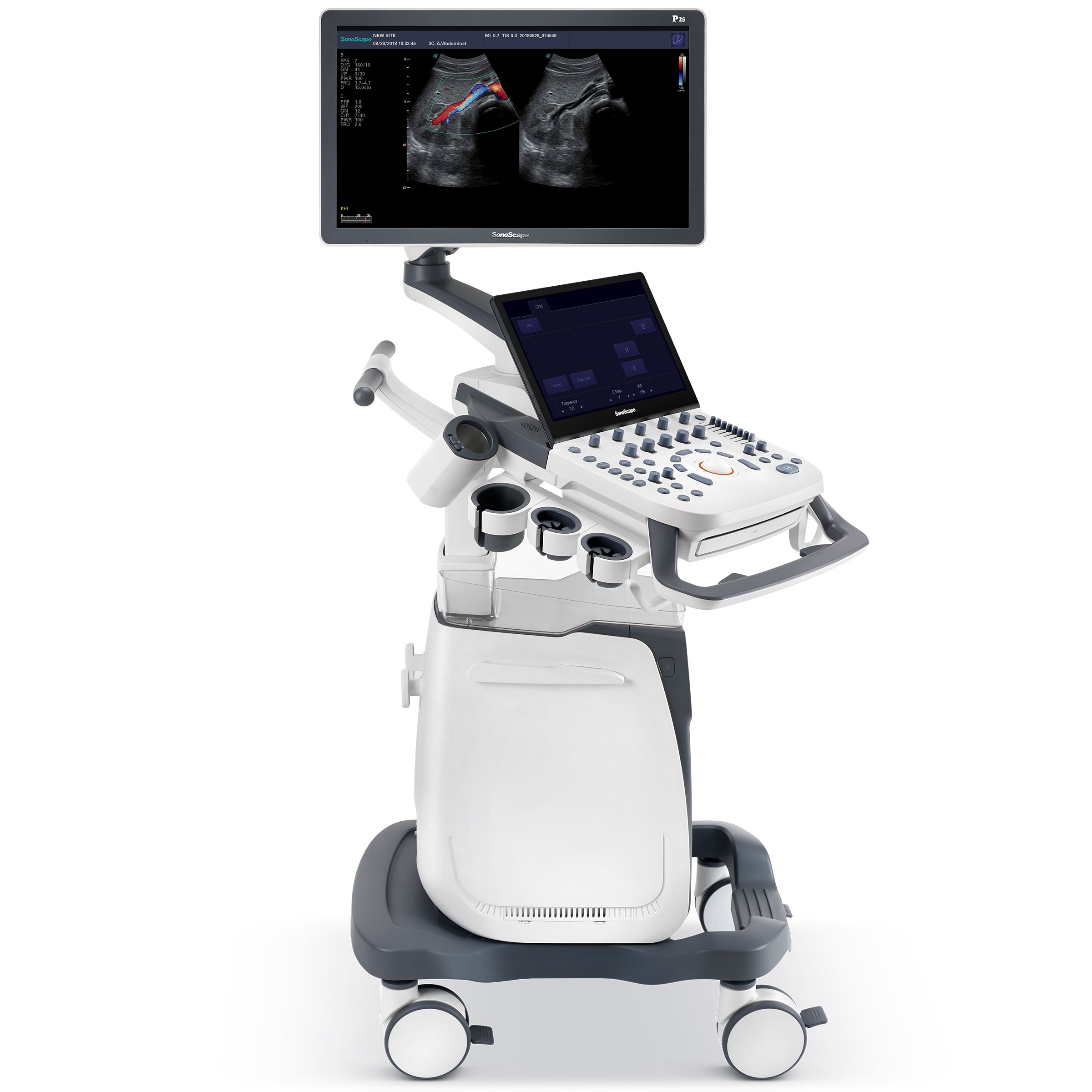
Based on the years of technical accumulation in the field of ultrasound, P25 is empowered by SonoScape’s cutting-edge technology to provide high-definition images to vastly increase diagnostic confidence. The sophisticated features and rich probe configurations provide a comprehensive solution to manage all-aspects of the daily clinical practice with ease and certainty.
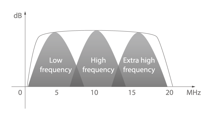
Broad Bandwidth Platform
The system ultra bandwidth and advanced probe technology enable better information collection. They provide higher resolution at depth with enhanced image quality for accurate diagnosis.
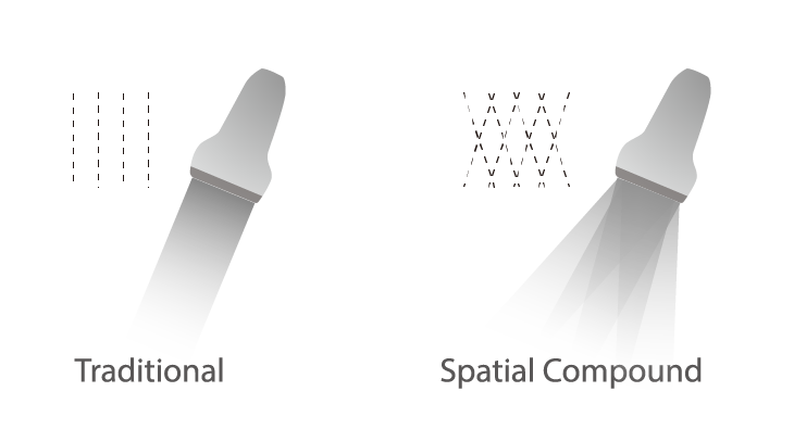
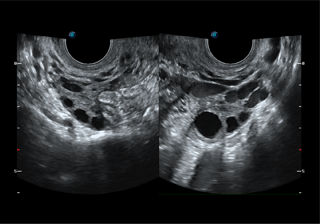
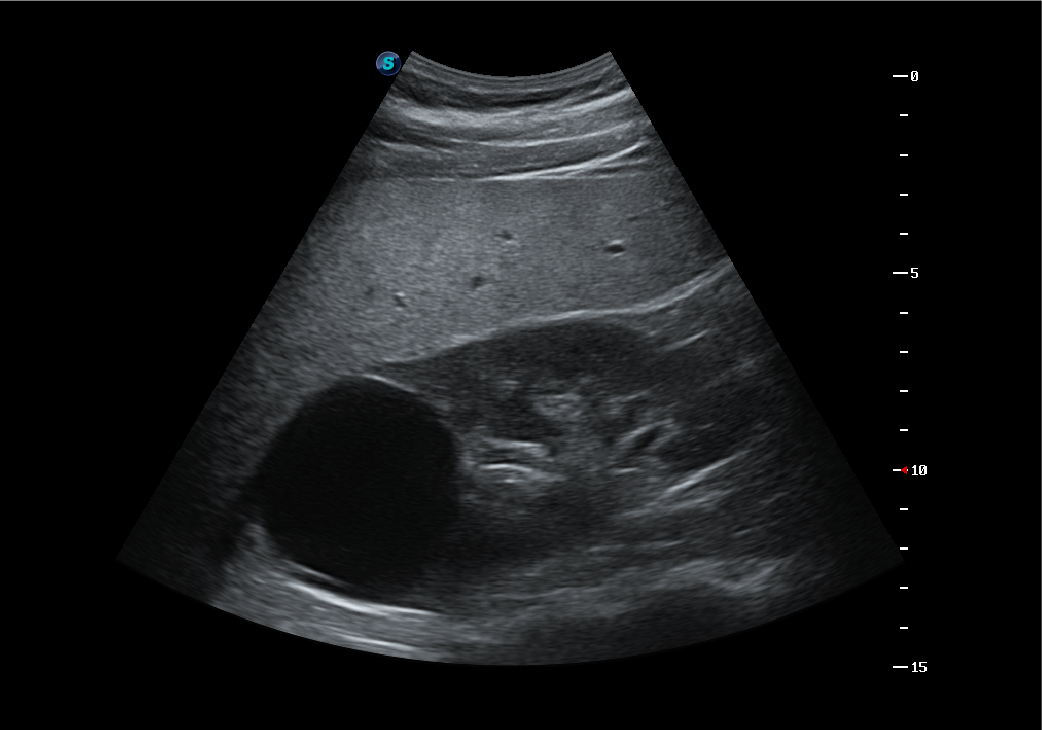
Spatial Compound Imaging
TheSpatial Compound Imaging utilizes several lines of sight for optimal contrast resolution, speckle reduction and border detection, with which P25 is ideal for superficial and abdominal imaging with better clarity and improved continuity of structures.
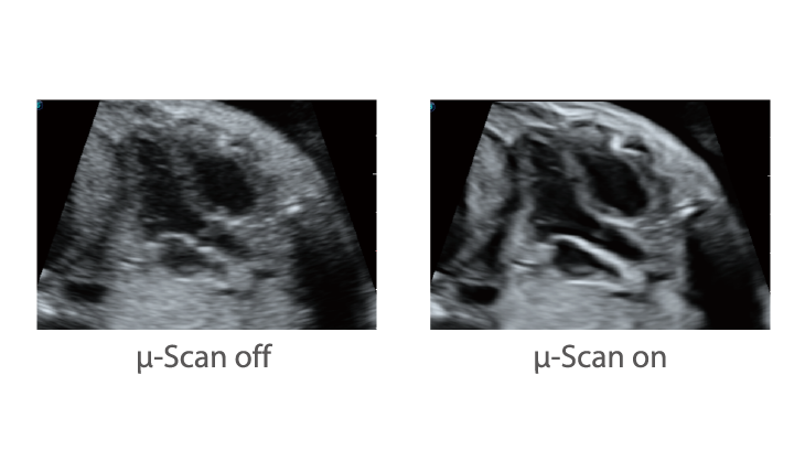
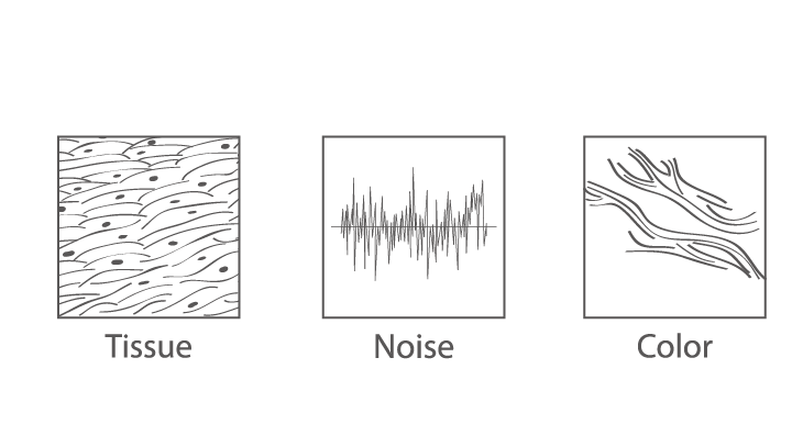
µ-Scan+
The new generation µ-Scan imaging technology gives you better image quality by reducing noise, improving signal strength and improving visualization.
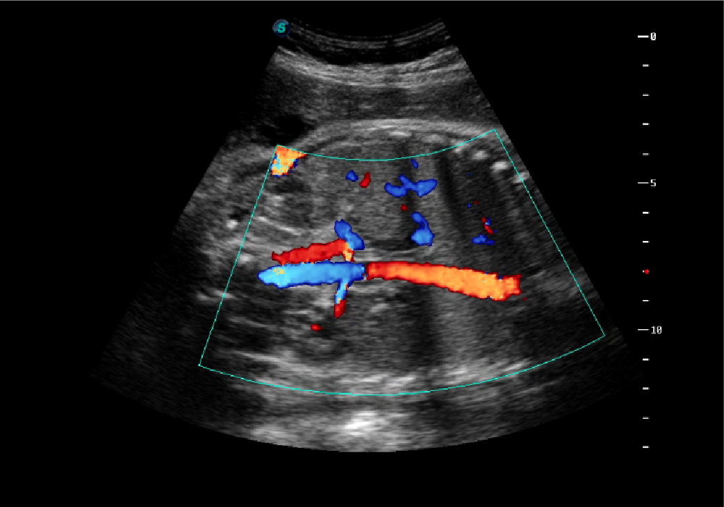
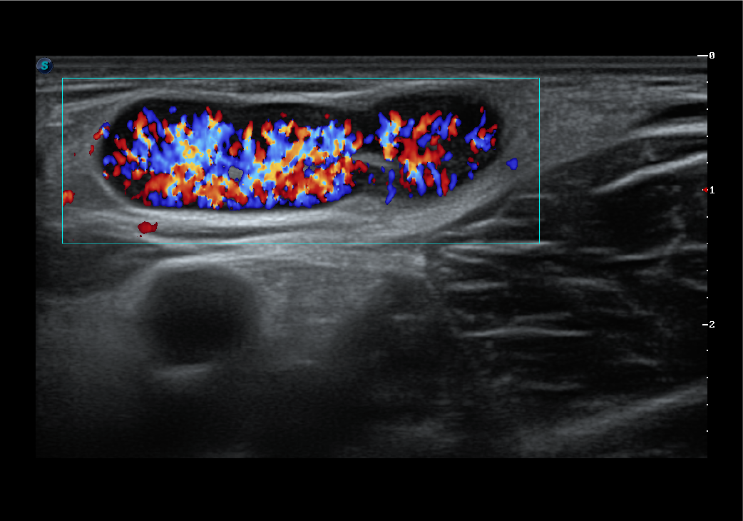
Dynamic Color
Dynamic color improves upon already existing color Doppler technologies for a clearer capture of color flow and detailed visualization of even tiny veins with lower velocities.
Extraordinary Images
To improve the diagnostic confidence of doctors, SonoScape endowed P25 excellent image quality so it can restore more details and information of anatomy structures with its comprehensive probes.
 |
 |
 |
 |
 |
 |
Comprehensive Features Cover Full-range Applications
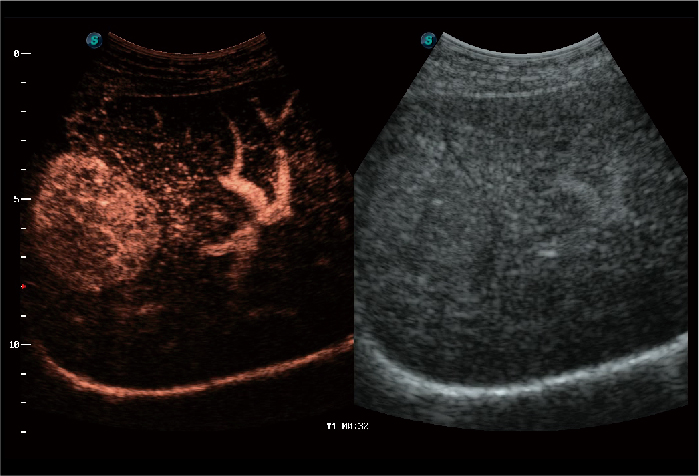 Contrast Imaging
The contrast agents provide a loud signal reflection, giving a more enhanced image of difficult-to-view blood flow. The Dynamic Acoustic Control feature of SonoScape Contrast Imaging provides image quality with a smaller agent dose.
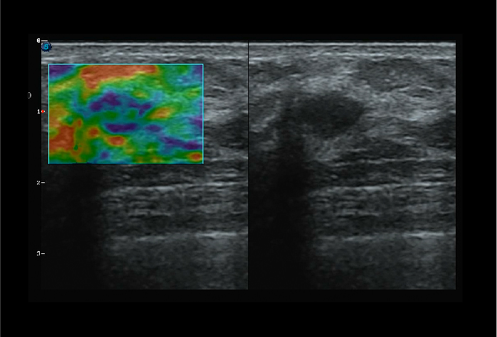 C-xlasto Imaging
C-xlasto Imaging enables comprehensive quantitative elastic analysis. It is supported by multiple probes to ensure good reproducibility and highly consistent quantitative elastic results.
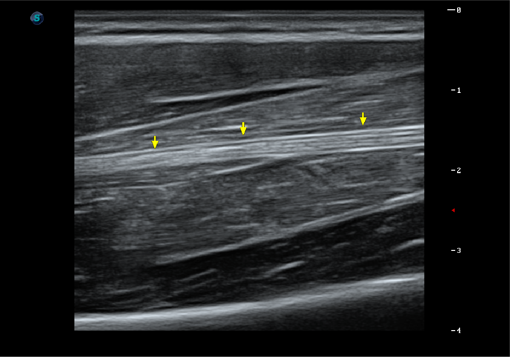
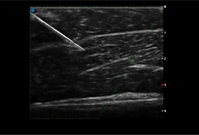 Vis-Needle
Vis-Needle is realized by ultrasound beam steering and deflection. It improves visualization of the needle shaft and needle tip in the tissue to minimize harm to the surrounding tissue, increasing the initial success rate and lowering the risk for needle puncture.
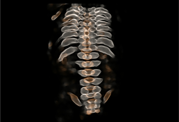 S-Live Silhouette
S-Live Silhouette is a unique transparent volume image for a more comprehensive internal and external view of the anatomy and provides more abundant diagnostic information for the clinic.
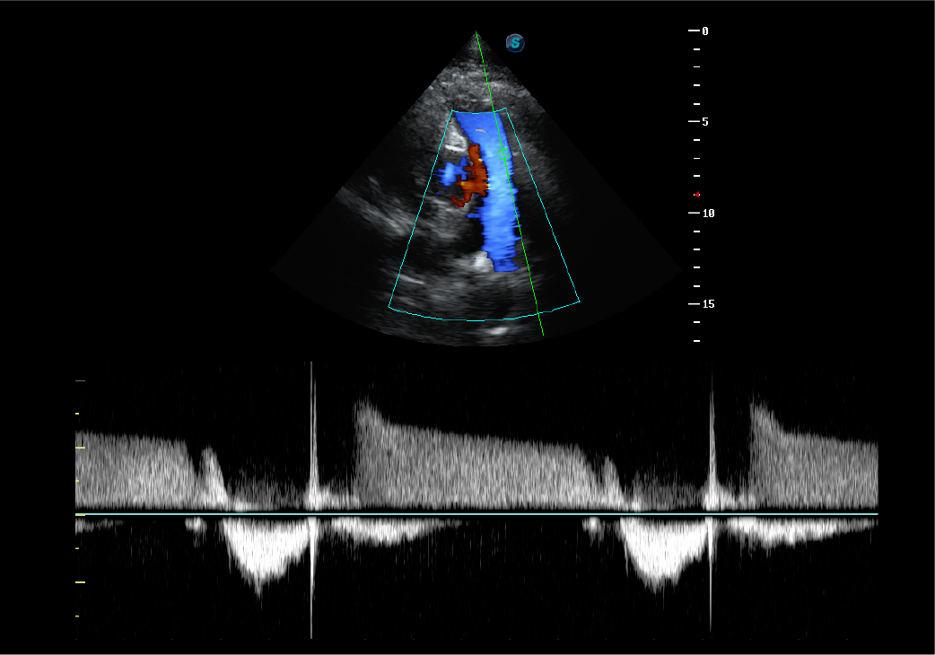
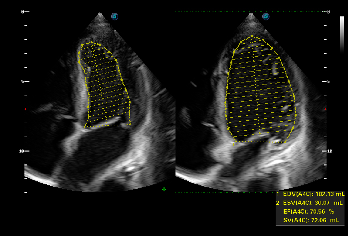 Auto EF
To recognize myocardial intima during the diastolic and systolic period and calculate ejection fraction automatically.
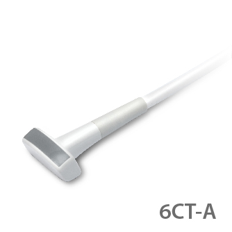
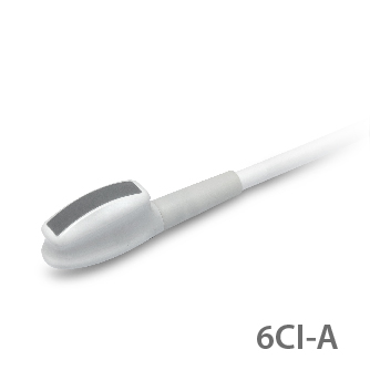
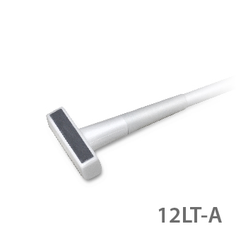
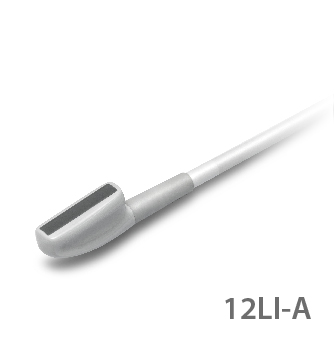
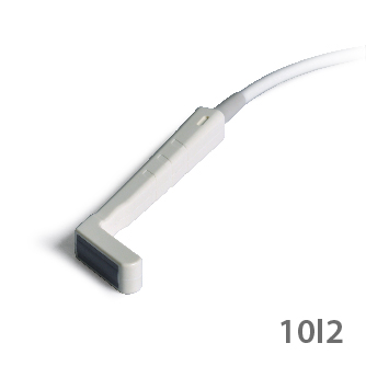
Specialty Probes for Specific Needs
P25 covers a wide range of intraoperative clinical needs with more than 5 specialty transducers that deliver the versatility to expand clinical offerings.