Intelligent Future Attainable

P60 Exp

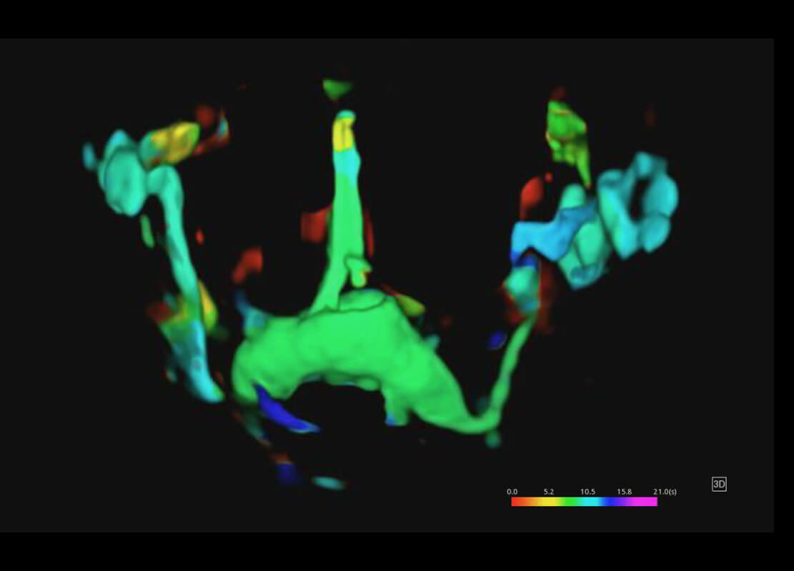
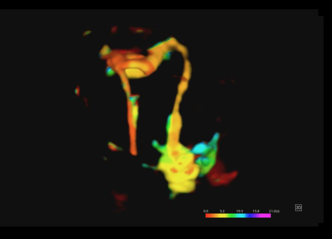
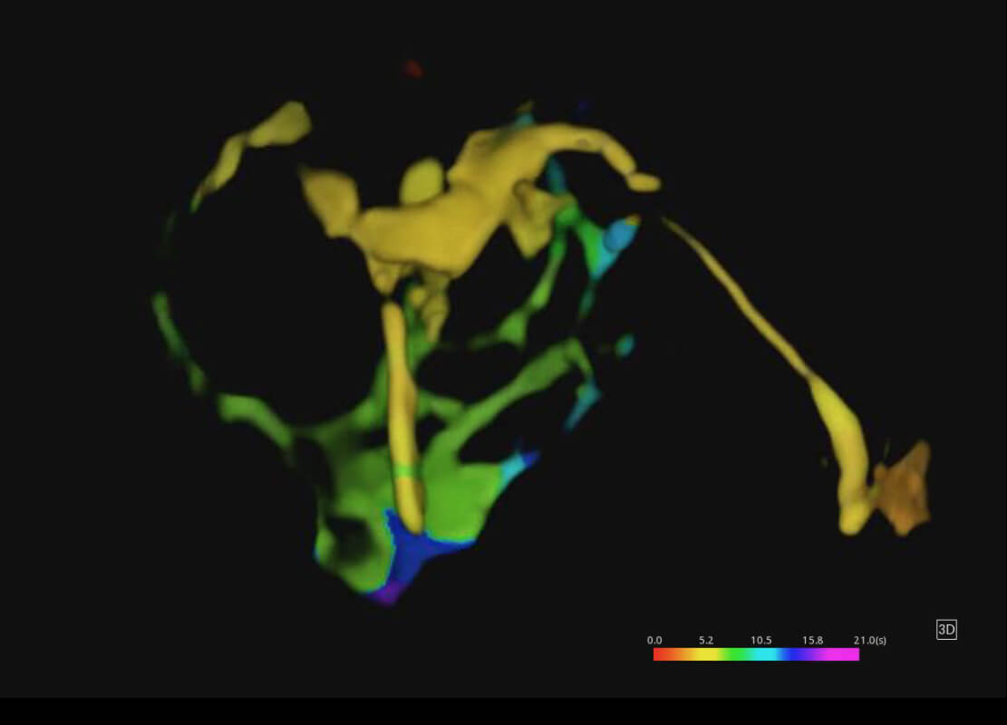
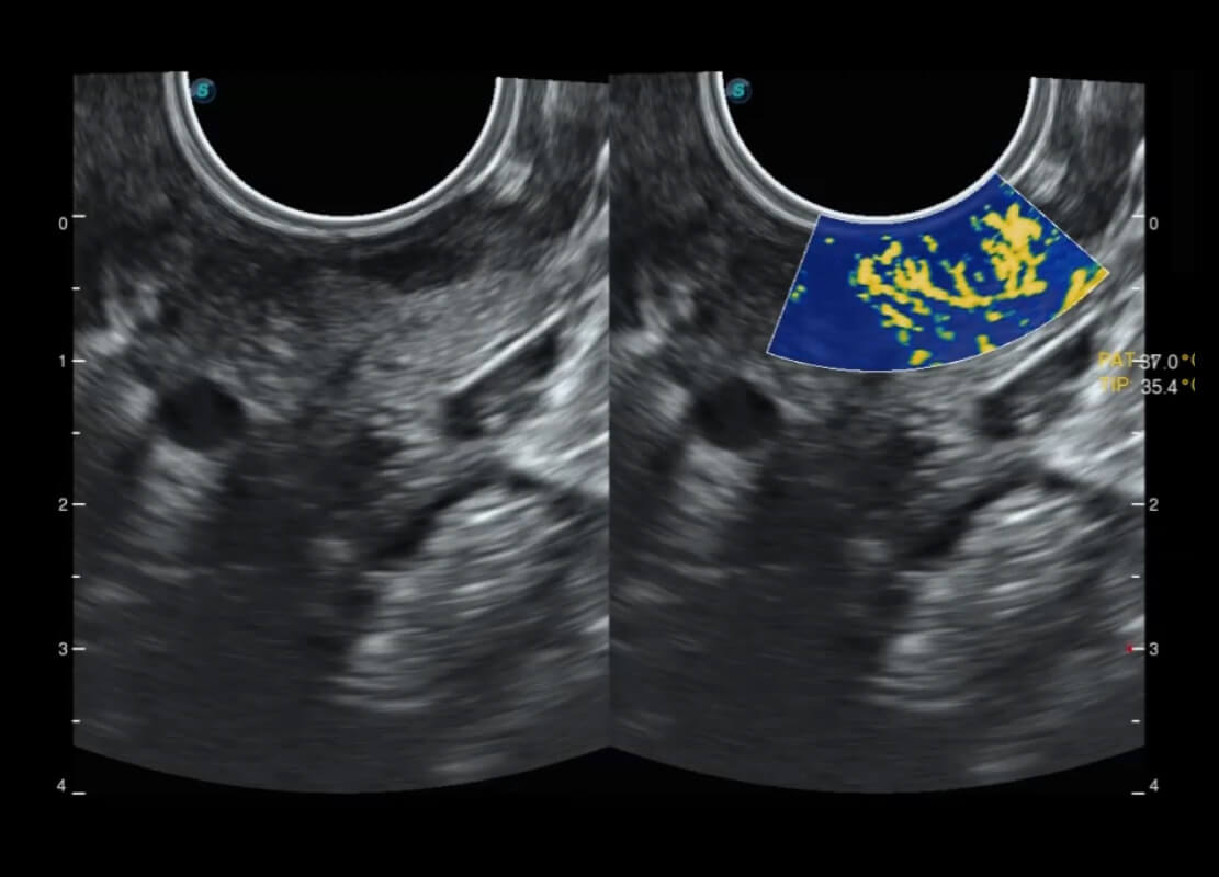
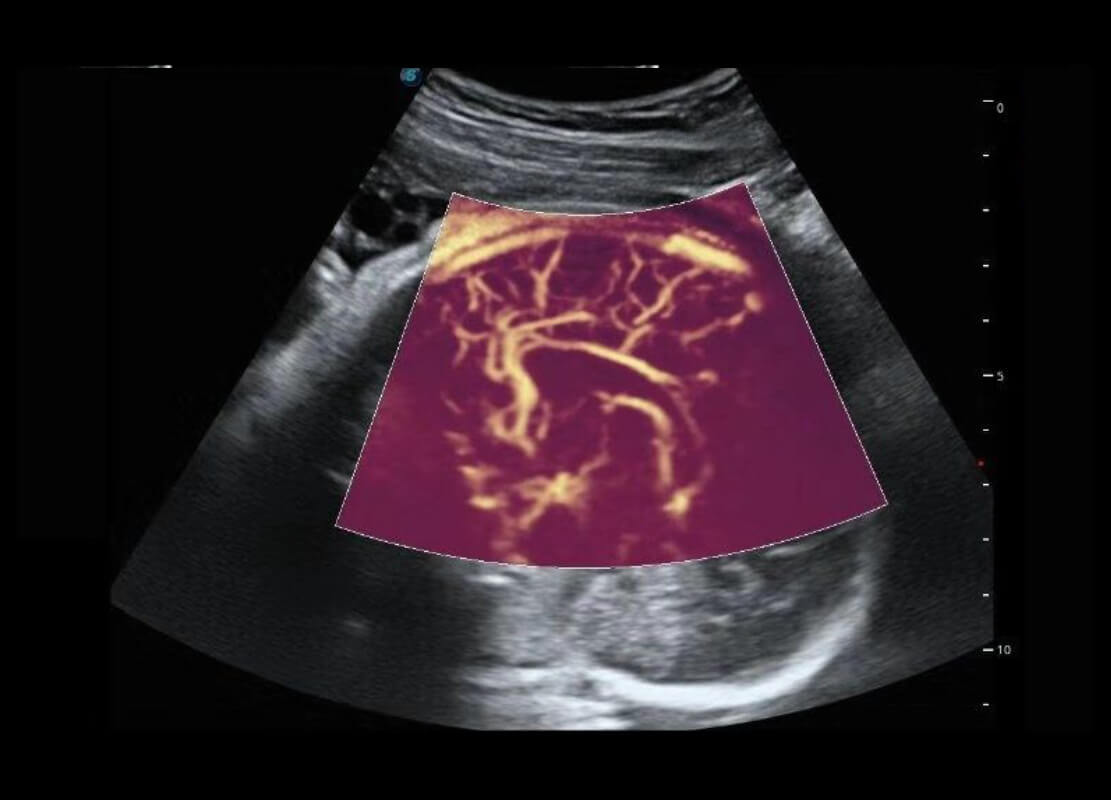
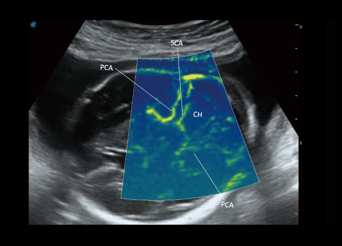
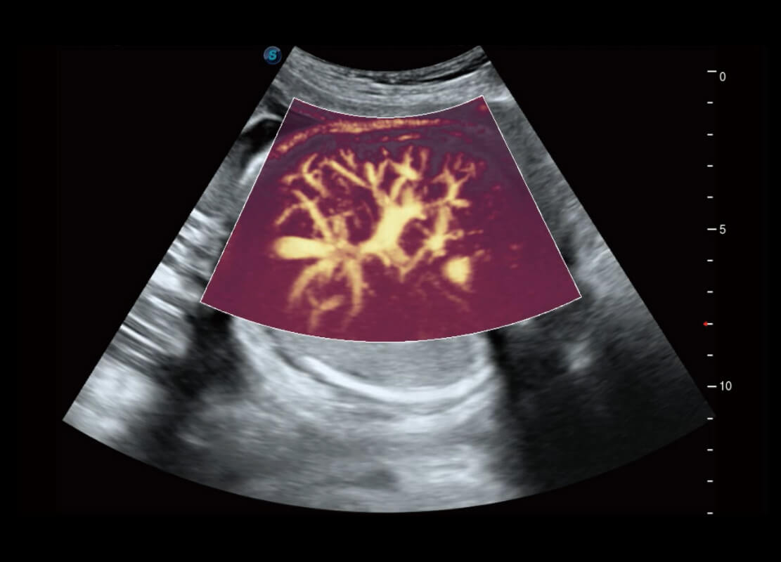
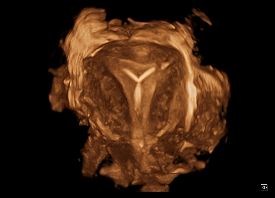
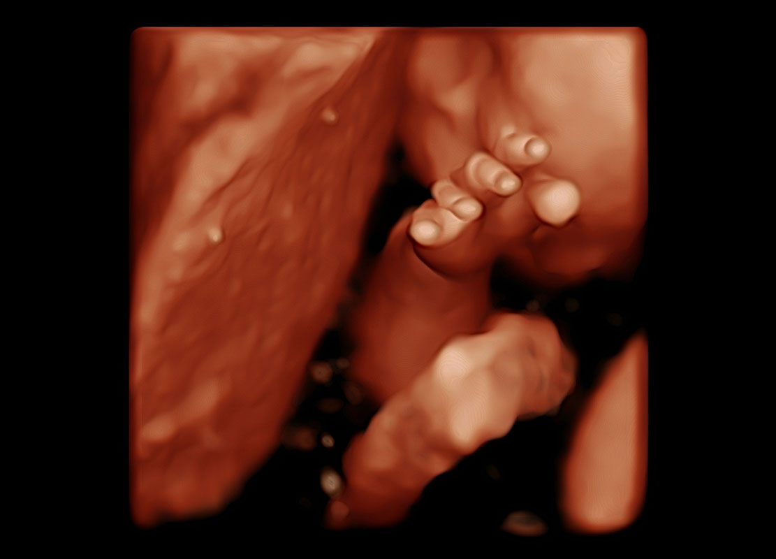
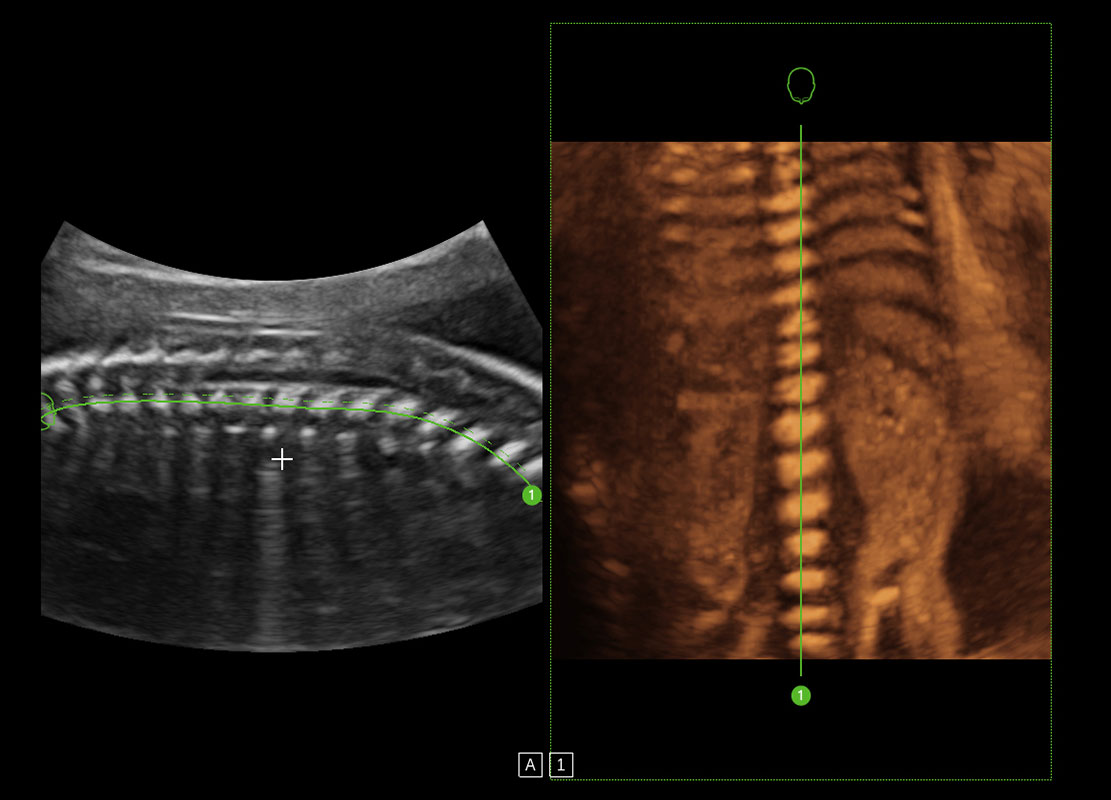
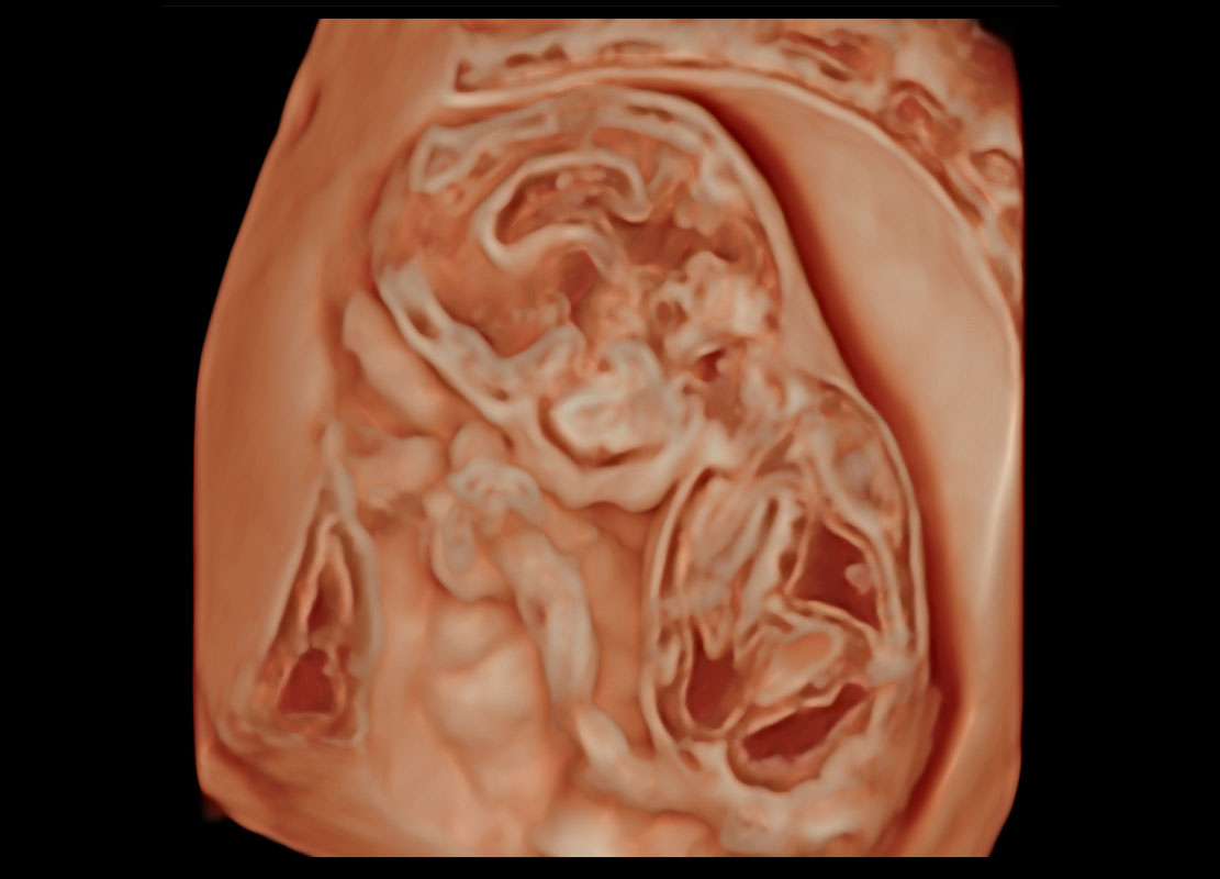
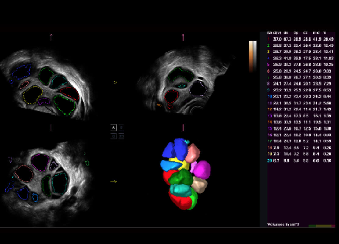
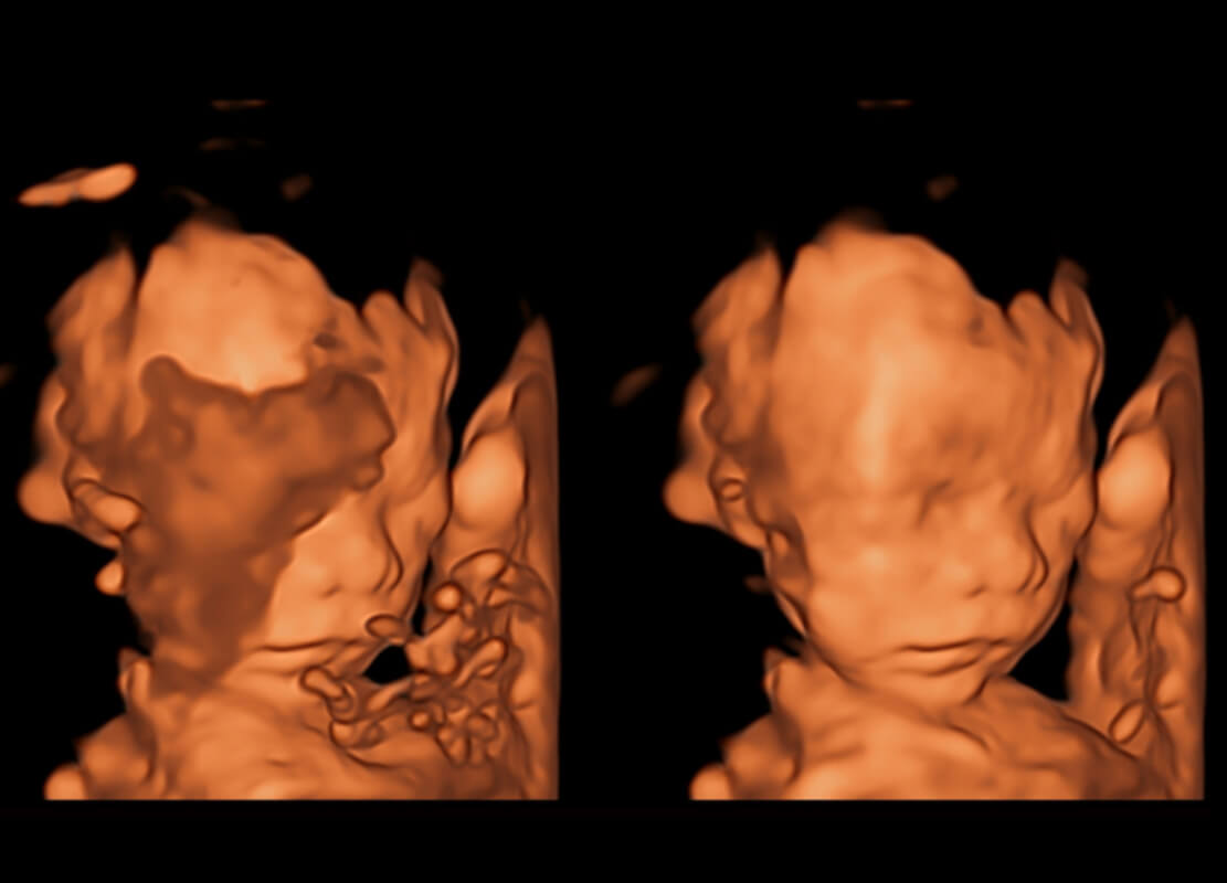
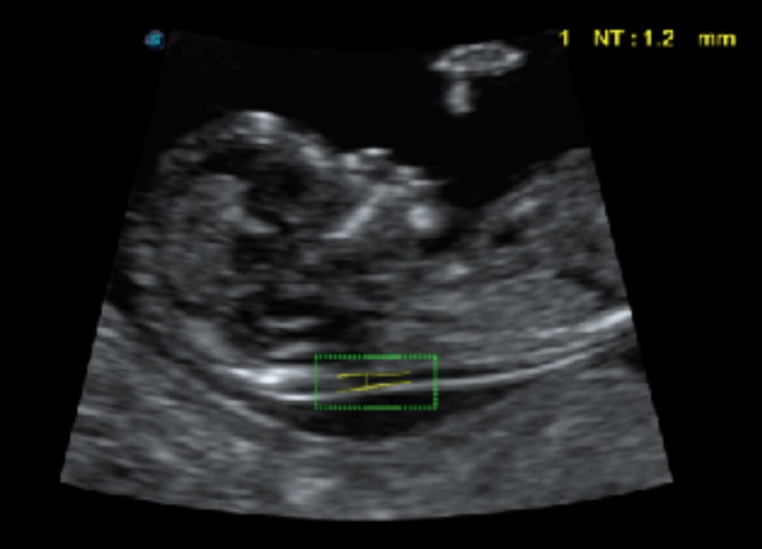
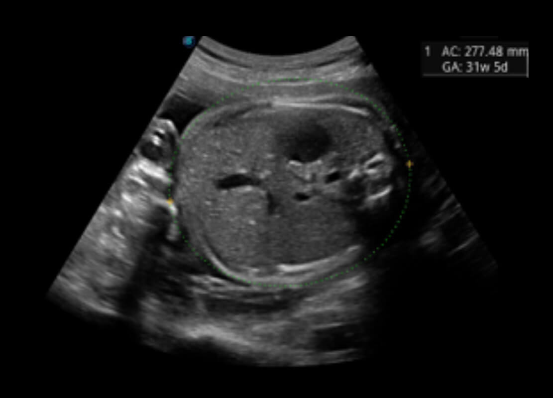
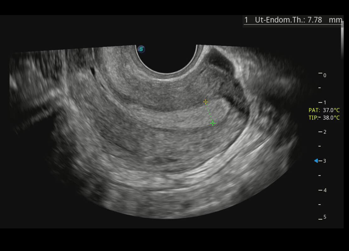
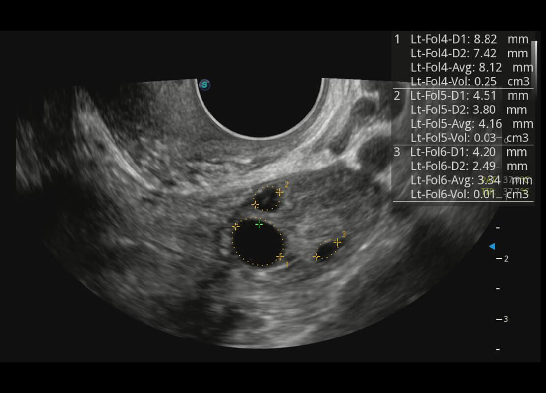
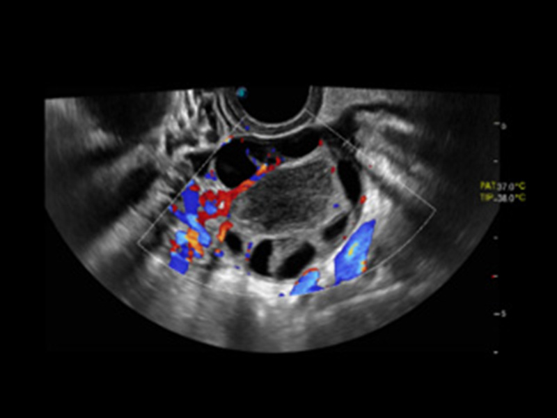
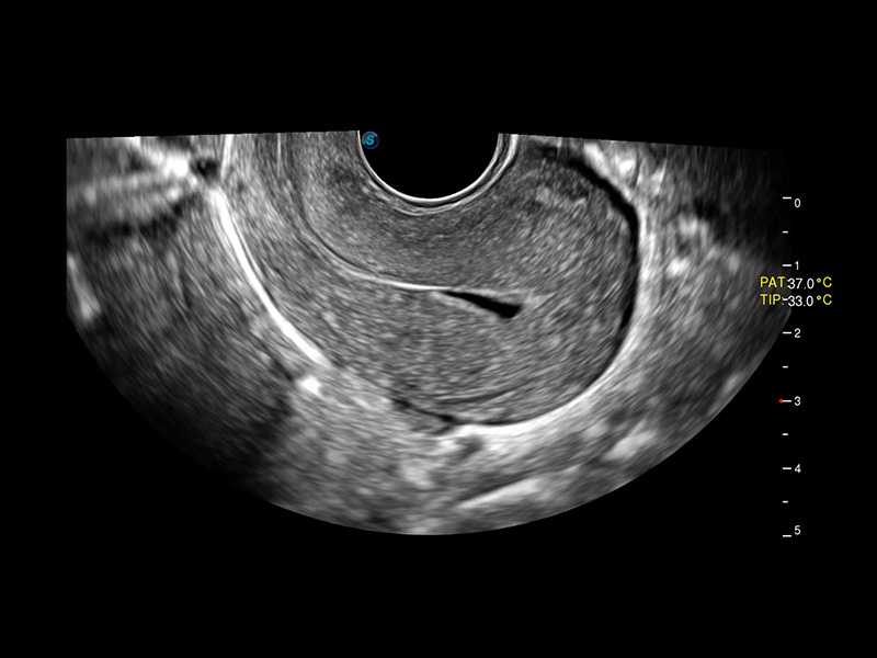
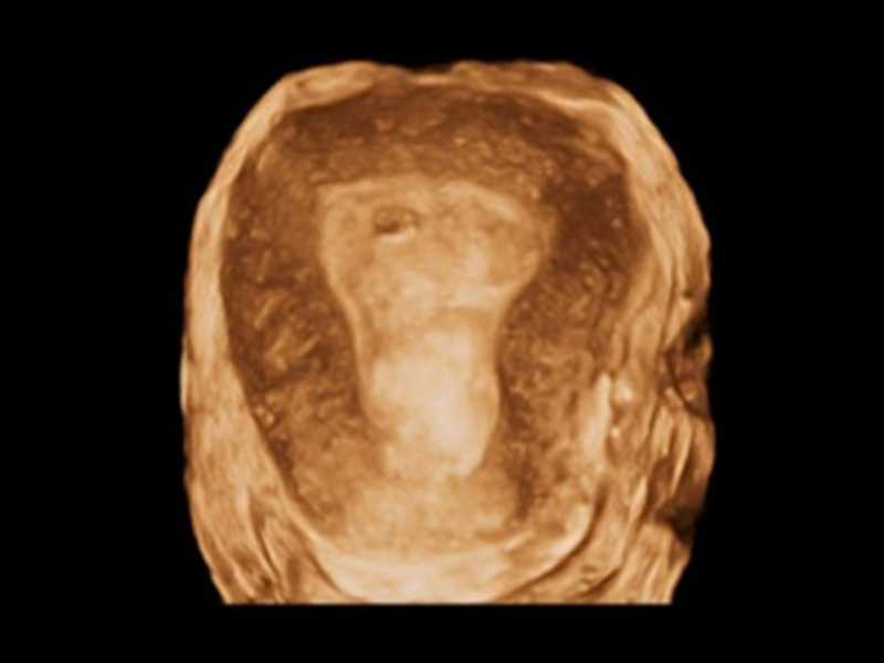
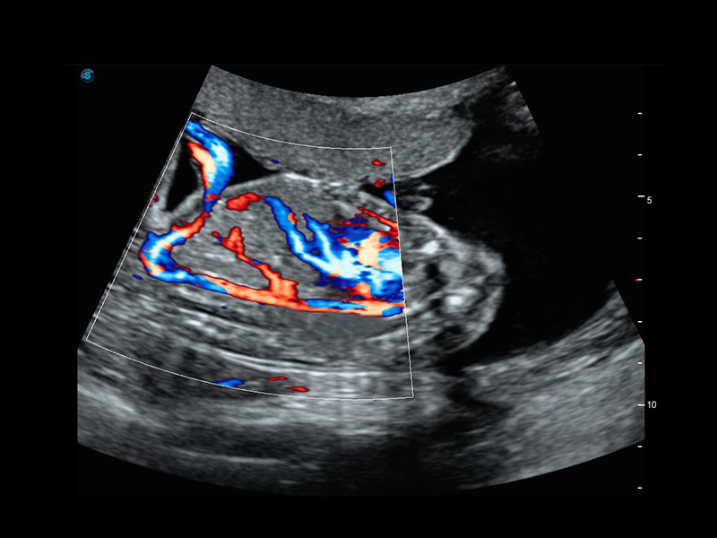
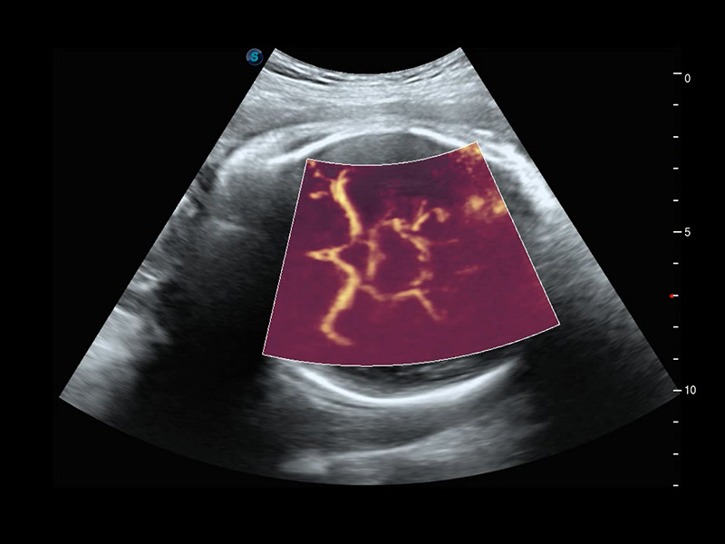
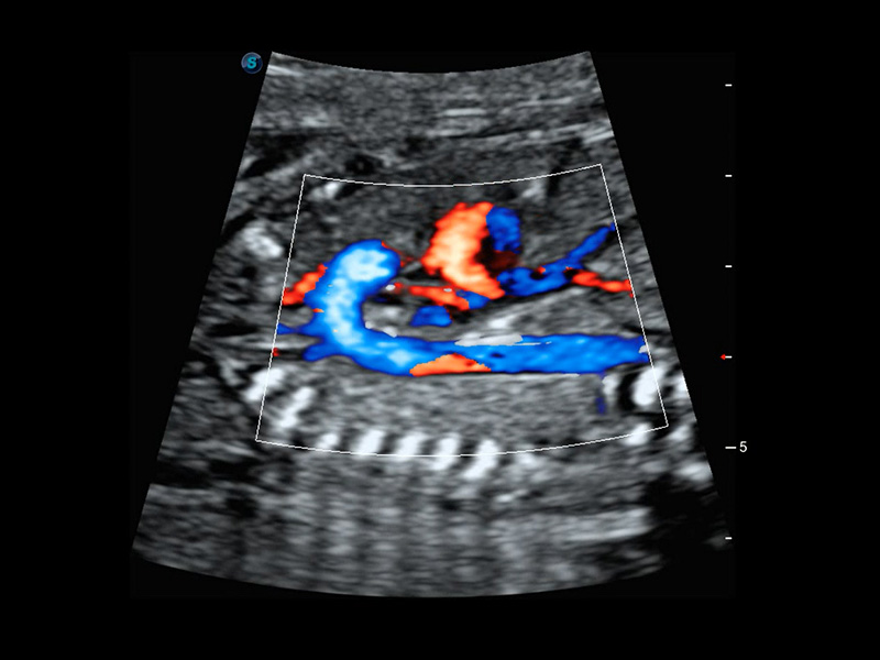
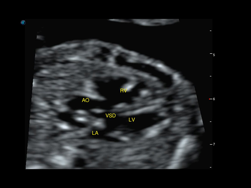
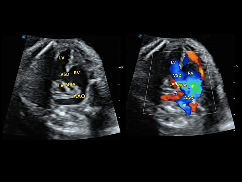
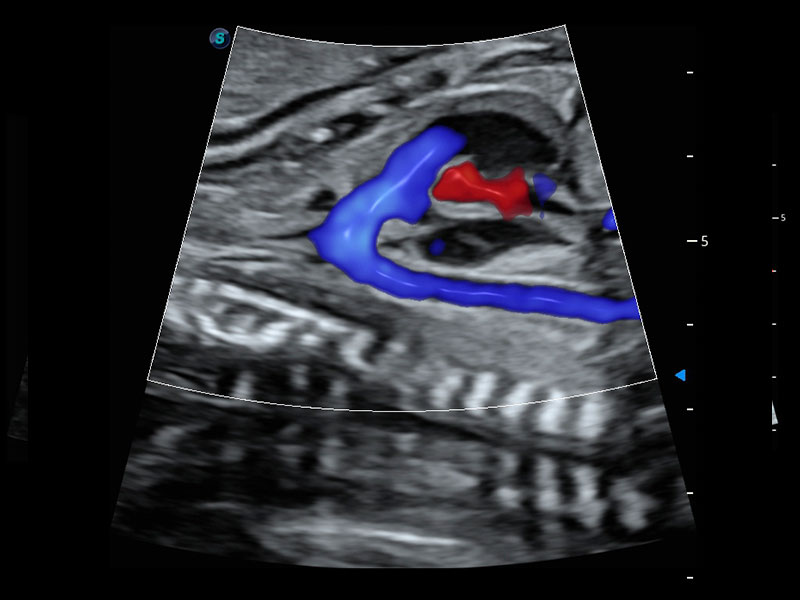
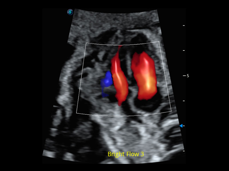
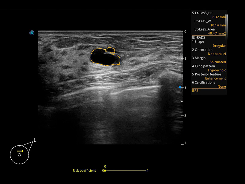
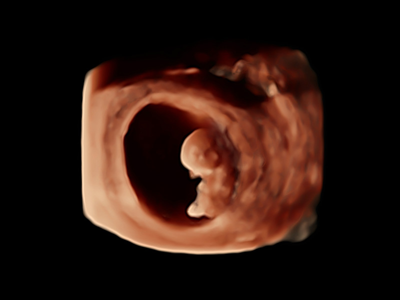
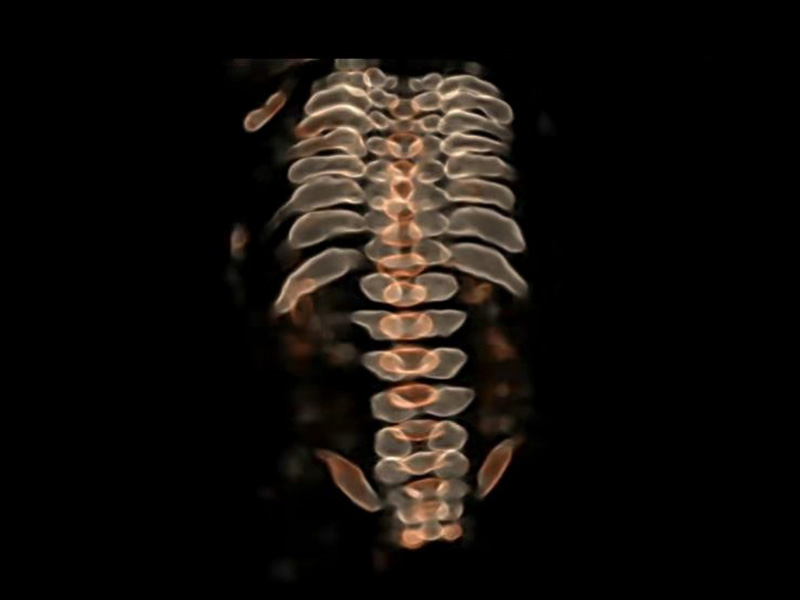
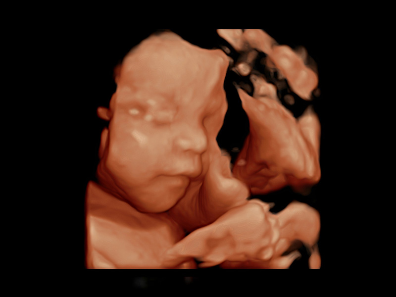
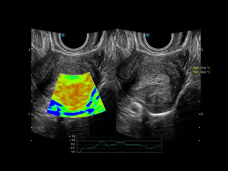
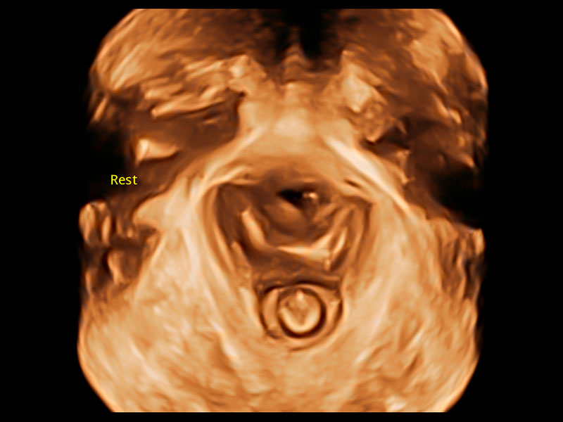
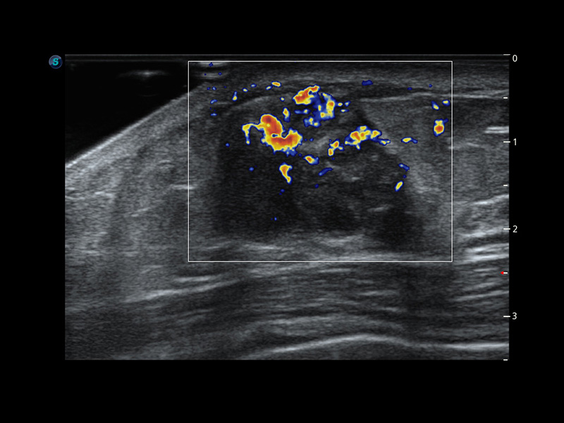
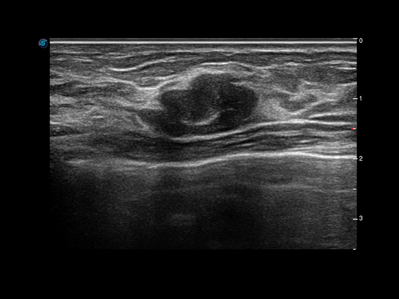
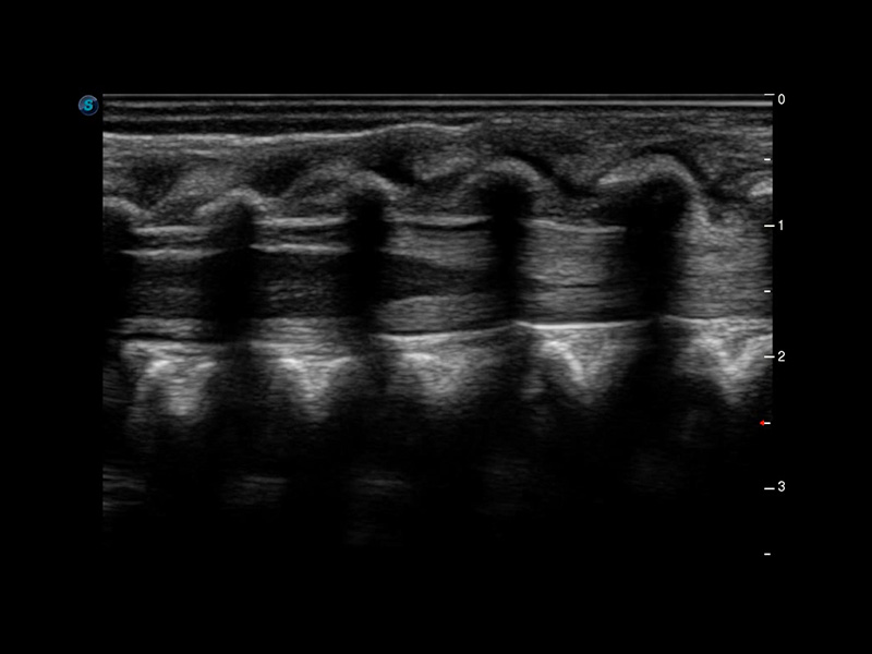
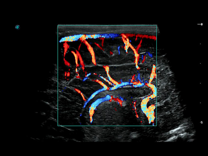
P60 Exp is designed to reform the practice at the frontend of women’s healthcare through continuous innovation. With a remarkable breakthrough in exceptional acoustic architecture and superior imaging capability, P60 Exp delivers true value to the detection and diagnosis of routine to complex cases in women’s health exams.

Wis+

An Artificial Intelligence Based Ultrasound Platform

  • Intelligent

  • Accurate

  • Efficient

Artificial Intelligence*

Brings unprecedented improvement on efficiency and accuracy

The adoption of AI on P60 Exp not only simplifies the workflow greatly, but also provides enhanced reproducibility and consistency in measurement.

1 OFD(HC): 87.03 mm
HC: 251.00 mm
GA: 27w 1d
HC/AC: 96.13 %

2 BPD: 70.56 mm
GA: 28 w 2d

S-Fetus(acq.)
& S-Fetus(meas.)

S-Fetus*

S-Fetus is a user-friendly tool allowing fully automatic and accurate detection of the most significant planes and frequently used measurements of fetal biometry. With a cine loop of the fetal head, S-Fetus can extract the standard planes and display the measurement results in one second, reducing the required keystrokes and working time remarkably by several times. It is designed to transform obstetric ultrasound examinations into a much cozier, faster and more delightful experience.

Lev.Hiat A-r: 16.33 cm2
Lev.Hiat H-r: 53.70 mm
Lev.Hiat W-r: 43.96 mm
Lt-LUG-r: 24.16 mm
Rt-LUG-r: 19.94 mm

S-Pelvic

S-Pelvic*

S-Pelvic is an advanced tool designed to reinvent the way clinicians evaluate Pelvic Floor Dysfunction (PFD). Due to highly intelligent capabilities, full automation of pelvic floor anatomy recognition, trace and measurement are now available, and can be achieved with one click at unprecedented ease. Moreover, S-Pelvic fulfills auto anterior compartment evaluation in 2D and auto levator hiatus evaluation in 3D/4D, and takes both rest and Valsalva maneuver into consideration, aiming to cover as many as possible steps and details in pelvic floor ultrasound and offer a comprehensive user experience.

* Due to regulatory reasons their future availability cannot be guaranteed.

4D HyCoSy with SPI

Elevated Capability Ready for More Challenges

Color coded Hysterosalpingo Contrast Sonography (HyCoSy) is a proprietary feature on P60 Exp that can clearly demonstrate the arrival time of contrast agents into different parts of the uterus, fallopian tubes and ovaries. As a result, clinicians are provided with strong and confident evidence to investigate tubal patency for subfertile female.

  • Fallopian Tube with 4D HyCoSy

  • Fallopian Tube with 4D HyCoSy with SPI

  • Fallopian Tube with 4D HyCoSy with SPI

  • Fallopian Tube with 4D HyCoSy with SPI

Micro F

Enables visualization for micro-vascularized structures

Micro F provides an innovative method to expand the range of visible flow in ultrasound, especially for visualizing slow flow tiny vessels. By adopting an advanced adaptive filter and accumulating temporal and spatial signals, Micro F can distinguish minute flow from overlaying tissue movement effectively, and depict hemodynamic with higher sensitivity and spatial resolution.

  • Cervical Poorly Differentiated Carcinoma with Micro F

  • Fetal Pericallosal Artery with Micro F

  • Fetal Unilateral Cerebellar Hypoplasia with Micro F

  • Fetal Hepatic Blood Flow with Micro F

Smart 3D/4D

Made Easy and Efficient

The new high density volume rendering engine of P60 Exp significantly optimizes signal processing and 3D rendering, making its volumetric performance outshines others in terms of high volume rates, exceptional details, and realistic effect. The comprehensive volume imaging suite extends your diagnostic capabilities into the next era of imaging with extraordinary image quality and streamlined workflow.

  • IUD with 3D Rendering

  • First Trimester Fetus with 3D S-Live Silhouette

  • Fetal Fingers with 3D S-Live

  • Fetal Spine with FreeVue and VCI

  • Fetal Inner Structure with S-Live Contour

Streamlined Workflow

Makes the interaction with ultrasound silky smooth

It is our commitment to make the user interaction with ultrasound as delightful and easy as possible through an ingenious design and diverse automation tools. P60 Exp is exactly a combination of both and enhances efficiency greatly by reducing keystrokes.

  • AVC Follicle

  • Auto Face

  • Auto NT

  • Auto OB (AC)

  • S-Endometrium

  • S-Follicle (2D)

Optimized Design

Inspires every ergonomic scanning

The design of P60 Exp stresses simplicity and compactness but makes no compromise to powerful performance. Height adjustable and lateral rotatable panel and an articulating monitor arm can basically satisfy any requirements under different scanning conditions.

  • 1
  • 2
  • 3
  • 4
  • 5
  • 21.5-inch LED Monitor

    P60 Exp features a large size 21.5-inch high definition LED monitor, providing excellent image display to users.

  • 13.3-inch Tilting Touch Screen

    13.3-inch touch screen allows users to browse and select functions with ease. A tilting design works for adjustment in terms of users’ needs.

  • User-friendly Layout

    Unique console design provides easy access to all kinds of common-used operation. Shortcut and customizable keys make it possible for users to tailor the workflow at their convenience.

  • Gel Warmer

    To ensure a comfortable patient experience, a gel warmer is available to be installed on the side of the control panel.

  • Built-in Battery

    A built-in battery supports P60 Exp to work for 2 hours without power supply, leaving users no worry about accidental pause and data loss due to power outage.

Image Gallery

* Technology and features are system/configuration dependent. SonoScape Medical reserves the right to change the above information and discontinue any product at any time without prior notification and will not be liable for any consequences resulting from the use of this publication.