C-Field+TM Architecture
Experience Innovation Redefined
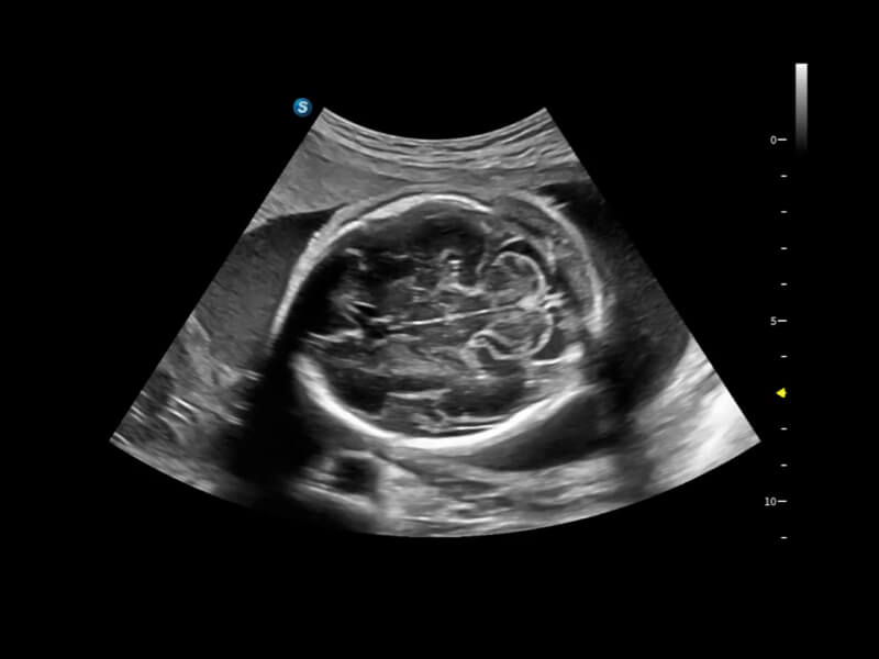
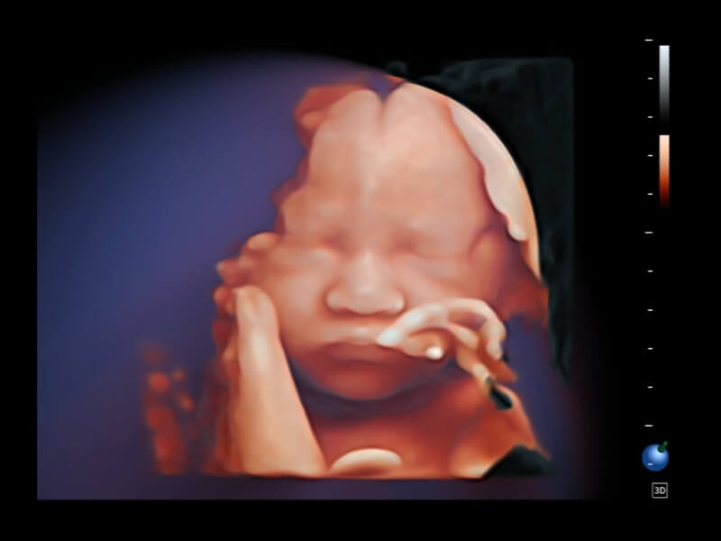
Experience Moments with Passionate Clarity
Discover the P80 ultrasound system, a cutting-edge solution designed to boost confidence and maximize efficiency in women’s health. With its advanced C-Field+TM architecture, the P80 delivers exceptional imaging quality and smart clinical solutions for obstetrics and gynecology, enhancing women's health protection.
Experience Innovation Redefined
Signal-to-Noise Ratio
Spatial & Contrast Resolution
Frame Rate
Improved Efficiency
Accelerated Computing
Boosted Processing
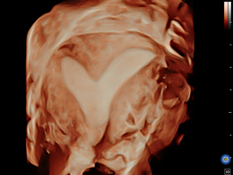
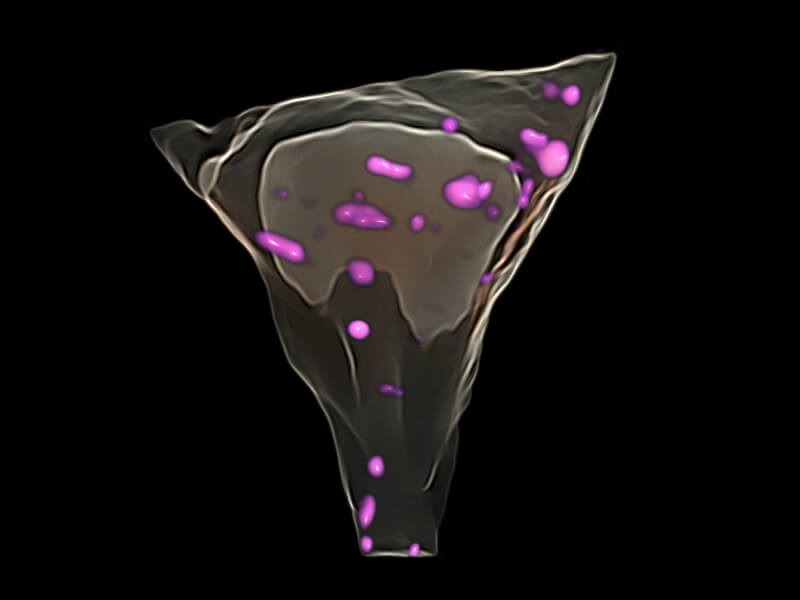
Seeing the anatomy in volume perspectives, with advanced assessment and intelligent scanning tools, these new perspectives provide clinicians with more information and help identify anomalies earlier than ever before.
The P80 revolutionizes patient care with streamlined workflow, remote access soutions, automatic probe activation, and an ergonomically optimized design, aiming to boost productivity and deliver superior care.
Probe Auto Wakeup
SonoSynch
SonoDrop
SonoAssistant
SonoHelp
SonoTransfer
Efficient Scanning
Interactive Connection
Educative Platform
Workflow Assistant
With intelligent motion boost sensor, new generation transducers support auto activation. Users can enjoy the convenience of multi-adjustable sensitivity levels tailored to their preferences, further enhancing examination flexibility and efficiency.
Real-time diagnosis interface and extra camera sharing, enabled by Sono-Synch, makes it possible to connect and adjust parameters by multi- ultrasounds and multi-smart devices in a remote distance at the same time, performing remote medical consultation and tutorial.
Onsite wireless data transmission tool allows to send patient file data independently, including ultrasound image, video and pdf report, clinicians can quickly share the scanning process in real-time and diagnosis result to patients and their families.
Helps streamline workflow while increasing standardization and reducing keystrokes and exam time by up to 50%.
An inspiring tutorial displaying probe placement, anatomy illustration and standard ultrasound image examples.
Allows to send the PDF report, image and cine data from ultrasound system to the shared folder on Windows computer for viewing.
Versatile & Elegant
Innovative & Effortless
Intuitive & Aesthetic
Smooth & Intelligent
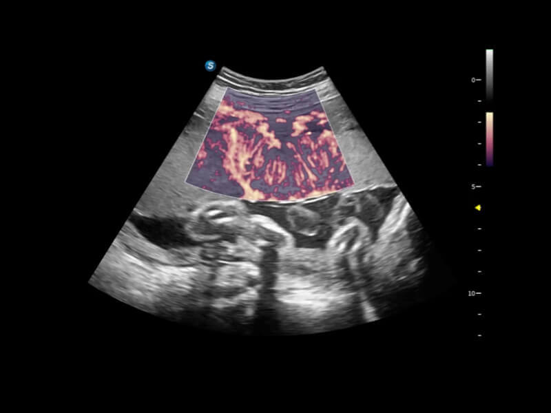
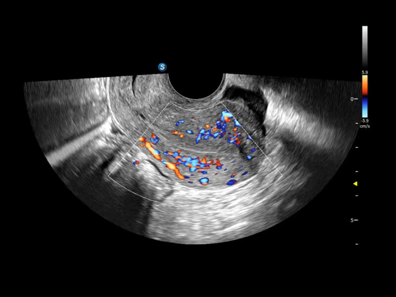
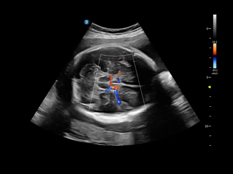
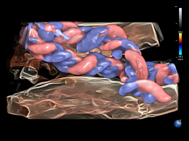
A wide variety of hemodynamic diagnostic techniques enable effective blood flow signal detection across different scenarios.
FHR enables the observation of microvascular structure within organs with enhanced sensitivity and improved resolution, ensuring the precision in representing the actual state of blood circulation.
Bright Flow offers 3D-like color Doppler flow visualization without requiring a volume transducer, thereby strengthening the boundary definition of vessel walls.
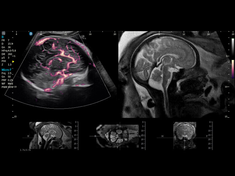
Micro F efficiently distinguishes subtle blood flow signals from overlaying tissue motion, enabling heightened sensitivity and spatial resolution in hemodynamic depiction.