S50
Reveal the Invisible
Reveal the Invisible

Adhering to its great image quality, SonoScape’s newly released S50 carries a comprehensive, upgraded platform, to become an epoch-making ultrasound system. It is equipped with powerful single crystal transducers, remarkable 4D functions and intelligent workflow, which greatly helps to meet a wide variety of general imaging needs. Outfitted with extraordinary software and hardware, S50 is a good balanced design, internally and externally. The quick response touch screen, mode switching and storage operations deliver a new standard with clarity and flexibility.
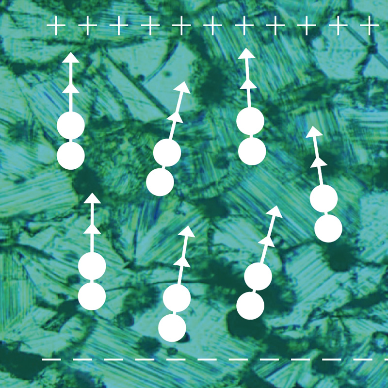
Single crystal: better penetration, higher resolution, greater sensitivity. Extremely clear image performance with unique imaging, optimized technology from SonoScape.
New platform leads to an efficient workflow and provides excellent human-machine interaction. It allows us to quickly respond to the needs of costumers as well as user defined settings.
SonoScape’s S50 provides various imaging software to fit different applications such as cardiology, radiology, OB/GYN, etc. With advanced features including cardiac and 4D packages, S50 can meet the extensive needs of clinical challenges of today and tomorrow.
SonoScape’s S50 is equipped with a wide band single crystal probe for abdominal and cardiac scanning, which can greatly improve signal to noise ratio, and acquire stunning images with better resolution and richer imaging detail. Compared with a conventional transducer, a single crystal probe has significantly improved acoustic energy conversion capacity, which means the probe has better performance as well as a longer working life.

The design of S50 took operational use into consideration, creating a comfortable diagnosing environment. Ergonomic design, excellent man-machine interaction and rapid response, makes S50 an intelligent scanning assistant for you, bringing improved efficiency and helping to prevent fatigue from multiple examinations.


By understanding that tissue stiffness varies depending on the type of tissue, we can use this to easily find abnormalities and tumors within soft tissue. When applying consistent and repeated pressure with the transducer on soft tissue the system can quickly calculate the Strain Ratio of the region and give an Intelligent Strain Curve for a recording of applied pressure to assist with diagnosis of the abnormality. Predominately used only with linear probes, SonoScape’s new transvaginal probe for gynecological is breaking the mold and expanding elastography applications to non-linear probes.
Contrast Imaging is the use of contrast agents that are small enough to go into the smallest of veins in the body and still provide a loud signal reflection for the ultrasound to pick up. Such a loud signal reflection allows the ultrasound to accurately display small veins in the body, blood profusion in different organs, and helps to measure blood flow. The S50 takes it a step farther by providing better resolution and deeper penetration, maximizing the short lifespan of the contrast agent bubbles by giving a clearer, more complete image of the desired organ.
SonoScape’s S50 is configured with a new imaging engine, which can significantly optimize image performance, especially for 3D/4D imaging with speed and convenience. Outstanding volume performance makes S50 outshine others on volume imaging, and dramatically enhances diagnostic confidence.
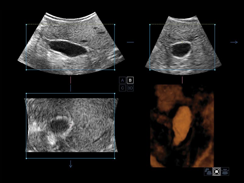
It provides more in-depth evaluation of vascular and cystic strictures creating a three-dimensional cast-like volume of the anatomy of interest.
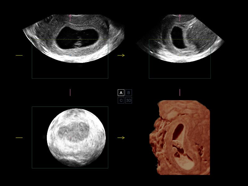
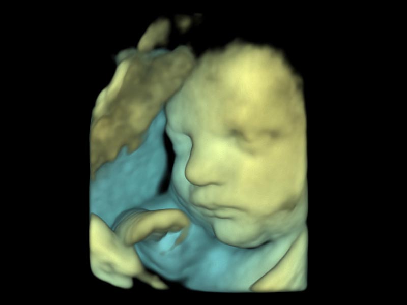
It allows for detailed visualization of subtle anatomical features, thereby enabling intuitive diagnosis on the real-time 3D images and enriching patient communication.
It can automatically display the near and foe distance relation from transducer to target, presented by smart designed color coding. It can help doctors to judge the spatial relationship on real-time 3D images.
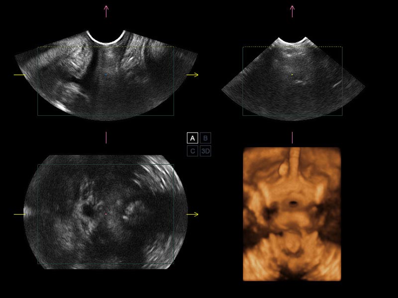
Transperineal 4D pelvic floor ultrasound can provide useful clinical values in assessing the vaginal delivery impact on the female anterior compartment, judging whether the pelvic organs are prolapsed or not and the extent, determining if the pelvic muscles were torn accurately.
Auto IMT is used when determining the level of vascular sclerosis present in the patient by automatically tracing and calculating the thickness of the carotid vessels.
Tissue Doppler Imaging allows you to quantitatively evaluate local myocardial movements and functions, with speed and strain/strain rate parameters.
Stress echocardiography is the combination of 2D echocardiography with a physical, pharmacological or electrical stress of the patient. It also provides users with report management tools such as configurable template editor, multiple loops to select one for storage, wall motion scoring, stress echo report, etc…

Fetus Spine of 4D

Uterus

Neonate Intracranial Color Flow

Kidney Color Flow

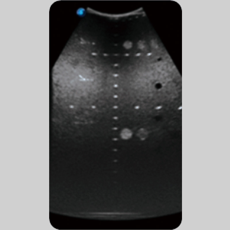
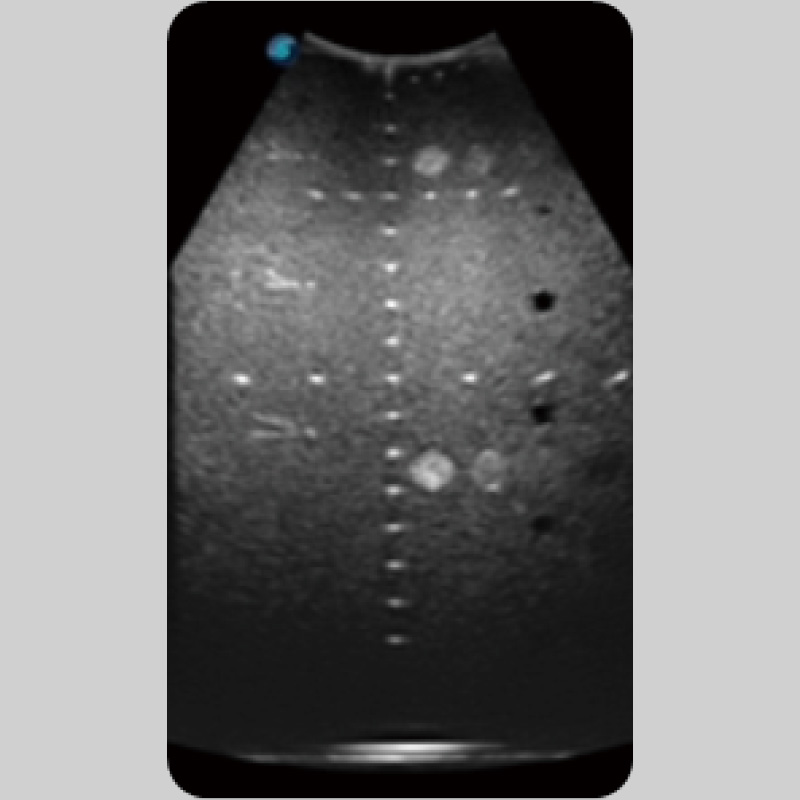
Liver

Slight Hydrocele of Testical

Cardiac

ICA Atherosclerotic Plaque PW

Pulmonary Regurgitation CW
SonoScape Medical Corp. stands as a prominent innovator in medical technology, specializing in ultrasound medical imaging, endoscopic diagnosis and treatment, minimally invasive surgery (MIS), and cardiovascular intervention solutions. Offering professional medical solutions and support in over 170 countries, SonoScape is driven by a passion for continuous innovation, unlocking life's potential and paving the way for boundless advancements in healthcare.
Copyright © SonoScape Medical Corp. All Rights Reserved. 粤ICP备20054866